Diagnostic Radiology
Mitch Goodsitt
Adjunct Professor, Nuclear Engineering and Radiological Sciences
Professor, Department of Radiology
[email protected]
Development of an Automated Spot Mammography Method for Improved X-Ray Imaging of Dense Breasts
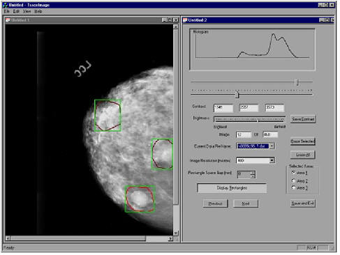
 One of the primary limitations of preset day x-ray mammography is the inability to optimally image regions of dense tissue within the breast. We are developing a novel technique to address this problem. The basic idea is to use computer aided diagnosis (CAD) methods to automatically detect any suspicious dense region within a whole breast digital mammogram and to take a second 3D stereo mammogram of only that region using more penetrating x-ray exposure and automated x-ray beam collimation. The resulting images are viewed on our in-house developed stereo display. Stereo mammography eliminates much of the superposition of tissues that occurs in conventional mammography. Thus, the new method should facilitate the detection and characterization of breast lesions that are located within dense tissue.
 Student developed graphical user interface that is used by radiologists to trace suspicious dense regions in mammograms for spot imaging. The radiologist-traced regions (in red) are compared with those determined by a computer aided detection (CAD) program.
Comparison of suspicious dense regions selected by the radiologist (in red) and the CAD program (green). The overlap between the areas is shown in yellow.
PI: Mitch Goodsitt, Ph.D., co-investigator: Heang-Ping Chan, Ph.D.
Development of Virtual Cursors for Measuring Depths in Stereo Mammography
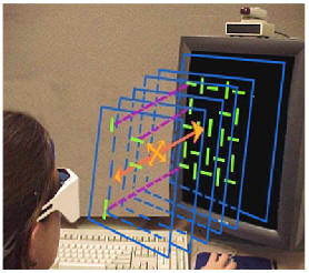
 2D cursors are routinely used to measure distances in digital radiographs and CT, ultrasound, and MR images. We have extended this concept to 3 dimensions and have developed virtual stereoscopic cursors that can be used to measure depths in stereo mammograms and stereo radiographs. We have performed observer studies and have determined that with the 3D cursor, it is possible to measure depths to an accuracy of about 2-mm. We are continuing our research in this area with the development of 3D pointers, 3D reference boxes, 3D translucent planes, etc.
PI: Heang-Ping Chan, Ph.D., co-investigator: Mitch Goodsitt, Ph.D.
Goodsitt MM, Chan, HP, Hadjiiski, L. Stereomammography: Evaluation of depth perception using a virtual 3D cursor. Medical Physics 2000; 27: 1305-1310.
Goodsitt MM, Chan HP, Darner KL and Hadjiiski LM. The Effects of Stereo Shift Angle, Geometric Magnification, and Display Zoom on Depth Measurements in Digital Stereomammography. Medical Physics 2002; 29: 2725-2734.
Development of a Device for a Combined X-Ray and Ultrasound Imaging of the Breast
Both x-rays and ultrasound have been found to be valuable in detecting and characterizing breast cancers. However, there is some uncertainty as to the correspondence between the lesions identified with the two modalities because the images are acquired with the breast in different geometries. We are developing a device for acquiring both x-ray and ultrasound images in the same geometry. The x-ray images are acquired using tomosynthesis methods to produce images of slices of the breast that can be directly compared with the ultrasound images. Advanced techniques such as 3D-grayscale ultrasound, 3D vascular ultrasound, elastography and computer aided diagnosis of the x-ray tomosynthesis and 3D ultrasound images will be explored.
PI: Paul Carson, Ph.D., co-PI: Mitch Goodsitt, Ph.D., with researchers at General Electric.
Analysis of Methods for Quantitative CT Measurement of Bone Mineral in Trauma Patients
Osteoporosis may be a contributing factor to the increased morbidity and mortality of elderly persons involved in motor vehicle accidents. To investigate this, we are proposing to measure the bone mineral densities of trauma patients. Such patients routinely undergo whole body CT scans. Quantitative CT is a method that is used to the measure bone mineral densities of patients. It is typically performed with the patient lying directly on top of a bone mineral calibration phantom. This is not possible or practical for trauma patients because of their condition and because they are lying on backboards. We have investigated alternative Quantitative CT calibration methods for this application using patient simulating phantoms and will continue this work with trauma patients.
PI: Stewart Wang, MD (General Surgery), co-investigator: Mitch Goodsitt, Ph.D.
Goodsitt MM, Christodoulou EG, Larson SC, Kazerooni EA. Assessment of calibration methods for estimating the bone mineral densities of trauma patients by quantitative computed tomography: An anthropomorphic phantom study. Academic Radiology 2001; 8: 822-834.
Nuclear Medicine
Robert A. Koeppe, Ph.D.
Professor of Radiology
Director PET Physics Section,
Division of Nuclear Medicine
[email protected]
Dr. Koeppe is responsible for overseeing the data acquisition and data analysis protocols for all research studies using positron emission tomography (PET). His area of expertise is kinetic analysis of dynamic PET data through the use of various compartmental modeling and parameter estimation techniques. His primary areas of research at present are two-fold. The first area is the development, testing, and optimization of data acquisition and analysis protocols for new PET radiopharmaceuticals that have been developed and brought on-line by the radiochemistry group at the University of Michigan. One current project that Dr. Koeppe is the principal investigator is to develop the radiotracer [18F]FEOBV (Fluoroethoxy-benzovesamicol) for the study of the vesicular acetylcholine transporter (VAChT). This project includes optimization of the radiochemical synthesis, validation studies in rodents and primates, and finally initial studies in humans, studying both normal control and patient subjects. Projects involving other new radiotracers follow similar steps. Dr. Koeppe’s second primary area of research, which as been supported by the Department of Energy for the past 5-6 years, is the development, implementation, and validation of dual-tracer PET methods. The goal of this work is to be able to assess multiple parameters of interest from a single-PET study that involves the injection of two different PET radiotracers. Since a PET scanner cannot distinguish between the different radiotracers, a 15-20 min offset in injection time and specialized kinetic analysis routines allow a study that normally would require approximately 4 hours to complete, to be finished within about 2 hours. This has a benefit both for the cost of the study, but also the convenience for the subject, and many patients are not able to tolerate the long scanning times required for multiple sequential PET studies.
Risk Analysis
Professor Ruth Weiner
Adjunct Professor, Nuclear Engineering and Radiological Sciences
(505) 284-8406
[email protected]
Links:
